Wednesday, January 26, 2011

Practical Applications And Potential Improvements of Cryogenics And Suspended Animation


Cryogenics
Every Wednesday, Michio Kaku will be answering reader questions about physics and futuristic science. If you have a question for Dr. Kaku, just post it in the comments section below and check back on Wednesdays to see if he answers it.
Today, Dr. Kaku addresses a question posed by Sassan K. Darian: What are the practical applications of cryogenics today, and what potential improvements can we expect 20 to 30 years down the line?


 The technology itself is almost 100 years old, but it took that long to get all the bugs out and create a new product that may revolutionize the media landscape. Other companies have slowly been implementing autostereoscopic technology into their devices. In 2009, Fujifilm released the FinePix Real 3D W1 digital camera which featured a built-in autostereoscopic LCD display and a twin-CCD/twin-lens system to capture your 3D images. (I have a chapter on this and other startling new technologies in my new book, Physics of the Future, also out in March.
In the future, perhaps all video and computer screens will employ a version of this technology. This could be the next big thing! Goodbye, clunky 3D glasses. Hello, the world of the Matrix.
Basically, these games use lenticular technology, the same technology used in novelty shops to create pictures that seem to fluctuate between two images (e.g. a person with eyes open, and then eyes closed). The key to these video games is the screen. The screen consists of many vertical lines, each one shaped somewhat like a prism. These vertical lines split an image in half, with one going to the left eye, and the other going to the right eye. Hence, each eye sees a slightly different image. When the brain puts these two images together, it creates the illusion of 3D. This technology dispenses totally with the glasses, since the image is split by the screen itself, not the glasses.
This development removes the main hurdle facing 3D technology. There are some drawbacks, which should be ironed out in the coming years, as TV, computer screens, and even flexible computerized wallpaper adopt this technology. You have to be in the sweet spot in order to get this full effect, about a foot behind the screen. If you are slightly outside the sweet spot, you experience ghosting, i.e. a slightly blurred image. (This is not so much a problem with video games, but it becomes more of a problem with TV screens that have to fill up a living room.)


Another challenge is software. Even though hardware may soon exist that will allow us to view 3D movies without glasses, Hollywood has to catch up and create movies for this new medium. In addition, creating software may be a bit expensive for this new technology. In lenticular technology, two separate images are broken up into many vertical strips, and then combined digitally, so they alternate, one strip after the next. Then the lenticular lens is placed right on top of these mixed strips. The prisms in these vertical lines in the lens separate this mixture into two separate images. This requires sophisticated technology. The integration of lenticular technology is just one step in the long road to creating a Matrix.
The 3DS does come pre-loaded with its own brand of built-in software including a Mii maker, Activity Log, Internet Browser, Backwards Compatibility, Camera and even the ability to download classic Nintendo games. One interesting feature is called the StreetPass™ which the 3DS website says "is the place where Mii™ characters meet and greet! When StreetPass™ communication is activated, you can exchange Mii data, recent gameplay info, and more with other Nintendo 3DS owners you pass on the street when your Nintendo 3DS system is in Sleep Mode. You'll then be able to see these Mii characters in the plaza the next time you start playing."

The next step, mentioned in my book, is to create Internet contact lenses that can shoot two 3D images directly into the retina of each eye. Imagine taking a final exam in the future using Interent contact lenses to download all the answers under the nose of the professor! Internet contact lenses could change everything, from our way of life to the world economy itself.


Sunday, January 23, 2011

The Basal Ganglia


Dr. C. George Boeree


Retrieved From:


Basal Ganglia


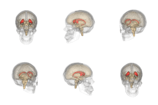
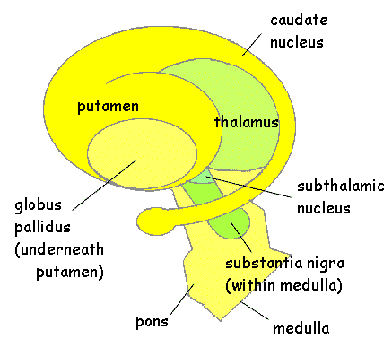
The basal ganglia are a collection of nuclei found on both sides of the thalamus, outside and above the limbic system, but below the cingulate gyrus and within the temporal lobes.  Although glutamate is the most common neurotransmitter here as everywhere in the brain, the inhibitory neurotransmitter GABA plays the most important role in the basal ganglia.

The largest group of these nuclei are called the corpus striatum ("striped body"), made up of the caudate nucleus ("tail"), the putamen ("shell"), the globus pallidus ("pale globe"), and the nucleus accumbens ("leaning").  All of these structures a double ones, one set on each side of the central septum.


The caudate begins just behind the frontal lobe and curves back towards the occipital lobe.  It sends its messages to the frontal lobe (especially the orbital cortex, just above the eyes), and appears to be responsible for informing us that something is not right and we should do something about it:  Wash your hands!  Lock your door!  As these examples are meant to suggest, obsessive compulsive disorder (OCD) is likely to involve an overactive caudate.  On the other hand, an underactive caudate may be involved in various disorders, such as ADD, depression, aspects of schizophrenia, and just plain lethargy.  It is also involved in PAP syndrome, a dramatic loss of motivation only recently discovered (see below).

The putamen lies just under and behind the front of the caudate.  It appears to be involved in coordinating automatic behaviors such as riding a bike, driving a car, or working on an assembly line.  Problems with the putamen may account for the symptoms of Tourette's syndrome.

The globus pallidus is located just inside the putamen, with an outer part and an inner part.  It receives inputs from the caudate and putamen and provides outputs to the substantia nigra (below).

The nucleus accumbens is a nucleus just below the previous nuclei.   It receives signals from the prefrontal cortex (via the ventral tegmental area) and sends other signals back there via the globus pallidus.  The inputs use dopamine, and many drugs are known to greatly increase these messages to the nucleus accumbens.

Another nucleus of the basal ganglia is the substantia nigra ("black substance").  Located in the upper portions of the midbrain, below the thalamus, it gets its color from neuromelanin, a close relative of the skin pigment.  One part (the pars compacta) uses dopamine neurons to send signals up to the striatum.  The exact function isn't known, but is believed to involve reward circuits.  Also, Parkinson's disease is due to the death of dopamine neurons here.

The other part of the substantia nigra (the pars reticulata) is mostly GABA neurons.  It's main known function is controlling eye movements.  It is also involved in Parkinson's, as well as epilepsy.


As you can see, quite a few serious problems are strongly associated with the basal ganglia.  Some, such as ADHD, Tourette's, obsessive-compulsive disorder, and schizophrenia, will be covered in other parts of this text.  Others are somewhat less psychological and more physical, but are still important....

Parkinson's disease

Parkinson's is characterized by tremor (shaking), rigid muscles, difficulty making quick, smooth movements, and difficulty standing and walking.  Many people also develop depression and anxiety and, later in life, problems with memory loss and dementia.

It usually develops late in life, but it can occur in younger people.  One well-known case is the actor Michael J. Fox.  It is very difficult for both the patient and his or her family.

Parkinson's is originates in the death of cells in the substantia nigra and the loss of dopamine and melanin produced by those cells.  It progresses to other parts of the basal ganglia and to the nerves that control the muscles, involving other neurotransmitters.  Possible causes or contributing factors include environmental toxins, head trauma, and genetics.

There are treatments available that slow the course of Parkinson's and alleviate the symptoms.  Most involve replacing or mimicking the lost dopamine and other neurotransmitters.  Unfortunately, the disease slowly progresses to where the treatments only work for a few hours at a time.  Parkinson's does not directly cause death and many patients live long lives with it.

Huntington's disease

Huntington's is characterized by loss of memory and odd jerking movements called chorea ("dance").  It is a hereditary disease (with a dominant gene) involving cell death in the caudate nucleus.  It usually starts in a person's 30s, but may start at any age.

 There is no cure, but there are treatments that can reduce the symptoms. It is fatal, although it is complications of the disease that usually cause death, rather than the disease itself.  Many Huntington's sufferers commit suicide.

Cerebral palsy

People with cerebral palsy have various motor problems, such as spasticity, paralysis, and even seizures.  Spasticity is where some muscles are constantly tight and so interfere with normal movement.  This is the reason for the unusual hand and arm positions most of us have seen in people with cerebral palsy.

It is apparently due to brain damage, usually sometime before birth.  Causes may include fetal infection, environmental toxins, or lack of oxygen.


(Although cerebral palsy tends to remain relatively stable throughout life, there is no cure and is very difficult to deal with for both the person and his or her family.)

With the aging process there are numerous complications, The person has to adjust to these complication.  Just ask me!  The "Umbrella of Cerebral Palsy" may stay stable, however, I must accept my deterioration with  the aging process!


PAP ( or Athymhormic) syndrome

PAP is characterized by an unusual lack of motivation.  A dramatic case was that of Mr. M, who, while drowning, simply failed to try to save himself, even though a good swimmer.

Damage to the caudate nucleus means that nothing carries any emotional significance anymore.  Drowning?  Don't be concerned.  People with PAP also ignore the usual social and moral motivations we all take for granted.  They don't quite "get" that their lack of action could have significant consequences.

Without the motivating influence of the basal ganglia, the frontal lobe simply stops planning for the future.  Oddly, they can still respond to external motivation, such as a loved one's request or an authority's command.

See the April 2005 Scientific American Mind article by Patrick Verstichal and Pascal Larrouy for more on PAP syndrome.

Saturday, January 22, 2011

UBC-VCH researchers find critical link between Down syndrome and Alzheimer's disease


Contact: Brian Lin
brian.lin@ubc.ca
604-822-2234
University of British Columbia


Retrieved From:


Link Between Down Syndrome and Alzhemer's


Researchers at the University of British Columbia and Vancouver Coastal Health Research Institute have discovered that the genetic mechanism which destroys brain cells is responsible for early development of Alzheimer's Disease in people with Down Syndrome and for development of Alzheimer's Disease in general population – providing a potential new target for drugs that could forestall dementia in people with either condition.
The research, led by Dr. Weihong Song, Canada Research Chair in Alzheimer's Disease and a professor of psychiatry in the UBC Faculty of Medicine, found that excessive production of a protein, called Regulator of Calcineurin 1 (RCAN1), sets in motion a chain reaction that kills neurons in the hippocampus and cortex in people with Down Syndrome and Alzheimer's Disease. The findings were published online recently in the Journal of Biological Chemistry.

"Neuronal death is the primary reason for the memory loss and other cognitive impairments of Alzheimer's Disease, and it's the main reason people with Down Syndrome develop Alzheimer's Disease long before most people, usually in their 30s," says Song, a member of the Brain Research Centre at UBC and the Vancouver Coastal Health Research Institute (VCHRI), and Director of Townsend Family Laboratories at UBC. "By looking for the common elements of both conditions, we were able to pinpoint how and why the deterioration occurs."

Alzheimer's Disease (AD) is the most common form of dementia, which usually affects people over age 60. The Alzheimer Society of Canada estimates that the disease affects more than 238,000 Canadians, and that by 2031 about 750,000 Canadians will suffer from AD and related dementias.

Down Syndrome (DS) is a congenital anomaly that includes developmental delays and learning disabilities. A 2002 report by the Public Health Agency of Canada said that about one in 800 Canadian newborns have the condition; the average lifespan for those with Down Syndrome is 49 years. People with DS have an extra copy of the gene that produces RCAN1, thus leading to its excess production. The resulting neuronal death – with symptoms that mirror those of AD patients – is one of the prime reasons for the shortened lifespan of people with DS.

The research team discovered that some AD patients have similarly elevated levels of the RCAN1 protein, despite having two copies of the responsible gene. It's still unknown why, though Dr. Song speculates that the gene's overexpression might be triggered by stroke, hypertension or the presence of a neurotoxic protein, called beta amyloid, that typically collects into clumps in the brains of people with AD – what he describes as a "vicious cycle" in which one destructive factor exacerbates another.

But now that the culprit gene and protein have been identified, "we can develop therapies that interfere with the gene's ability to produce that protein, and hopefully short-circuit the destruction of brain cells," Dr. Song says.
###
The research was supported by the Canadian Institutes of Health Research, the Jack Brown and Family Alzheimer's Research Foundation, the Michael Smith Foundation for Health Research and the National Natural Science Foundation of China.
Townsend Family Laboratories was established at The University of British Columbia with a donation of $7.5 million from the David Townsend Family. The research centre is dedicated to integrating the basic and clinical research for finding the underlying mechanism and novel diagnostic biomarkers for Alzheimer's Disease and developing interventions to prevent and treat this devastating disease.

The UBC Faculty of Medicine provides innovative programs in the health and life sciences, teaching students at the undergraduate, graduate and postgraduate levels, and generates more than $200 million in research funding each year. In 2007/08, out of the total UBC research endeavour, 53 per cent, or $247 million, came from academic and clinical teams in the Faculty of Medicine. For more information, visit www.med.ubc.ca.

The Brain Research Centre comprises more than 200 investigators with multidisciplinary expertise in neuroscience research ranging from the test tube, to the bedside, to industrial spin-offs. The centre is a partnership of UBC and VCH Research Institute. For more information, visit www.brain.ubc.ca.

Vancouver Coastal Health Research Institute (VCHRI) is the research body of Vancouver Coastal Health Authority, which includes BC's largest academic and teaching health sciences centres: VGH, UBC Hospital, and GF Strong Rehabilitation Centre. In academic partnership with the University of British Columbia, VCHRI brings innovation and discovery to patient care, advancing healthier lives in healthy communities across British Columbia, Canada, and beyond. www.vchri.ca.

Genetic Code for Form of Pancreatic Cancer Cracked


Retrieved From:

Pancreatic Cancer

ScienceDaily (Jan. 21, 2011) — Scientists at Johns Hopkins have deciphered the genetic code for a type of pancreatic cancer, called neuroendocrine or islet cell tumors. The work, described online in the Jan. 20 issue of Science Express, shows that patients whose tumors have certain coding "mistakes" live twice as long as those without them.


"One of the most significant things we learned is that each patient with this kind of rare cancer has a unique genetic code that predicts how aggressive the disease is and how sensitive it is to specific treatments," says Nickolas Papadopoulos, Ph.D., associate professor at the Johns Hopkins Kimmel Cancer Center and director of translational genetics at Hopkins' Ludwig Center. "What this tells us is that it may be more useful to classify cancers by gene type rather than only by organ or cell type."

Pancreatic neuroendocrine cancers account for about five percent of all pancreatic cancers. Some of these tumors produce hormones that have noticeable effects on the body, including variations in blood sugar levels, weight gain, and skin rashes while others have no such hormone "signal."

In contrast, hormone-free tumors grow silently in the pancreas, and "many are difficult to distinguish from other pancreatic cancer types," according to Ralph Hruban, M.D., professor of pathology and oncology, and director of the Sol Goldman Pancreatic Cancer Research Center at Johns Hopkins. For the new study, the team investigated non-hormonal pancreatic neuroendocrine tumors in 68 men and women. Patients whose tumors had mutations in three genes -- MEN-1, DAXX and ATRX -- lived at least 10 years after diagnosis, while more than 60 percent of patients whose tumors lacked these mutations died within five years of diagnosis.

The Johns Hopkins team, which previously mapped six other cancer types, used automated tools to create a genetic "map" that provides clues to how tumors develop, grow and spread.

Within the code are individual chemicals called nucleotides, which pair together in a pre-programmed fashion to build DNA and, in turn, a genome. Combinations of these nucleotide letters form genes, which provide instructions that guide cell activity. Changes in the nucleotide pairs, called mutations, can create coding errors that transform a normal cell into a cancerous one.

In the first set of experiments, the Johns Hopkins scientists sequenced nearly all protein-encoding genes in 10 of the 68 samples of pancreatic neuroendocrine tumors and compared these sequences with normal DNA from each patient to identify tumor-specific changes or mutations. In another set of experiments, the investigators searched through the remaining 58 pancreatic neuroendocrine tumors to determine how often these mutated genes appeared. The most prevalent mutation, in the MEN-1 gene, occurred in more than 44 percent of all 68 tumors. MEN-1, which has been previously linked to many cancers, creates proteins that regulate how long strands of DNA are twisted and shaped into dense packets that open and close depending on when genes need to be activated. Such a process is regulated by proteins and chemicals that operate outside of genes, termed "epigenetic" by scientists.

Two other commonly mutated genes, DAXX and ATRX, which had not previously been linked to cancer, also have epigenetic effects on how DNA is read. Of the samples studied, mutations in DAXX and ATRX were found in 25 percent and 17.6 percent, respectively. The proteins made by these two genes interact with specific portions of DNA to alter how its chemical letters are read.

"To effectively detect and kill cancers, it may be important to develop new diagnostics and therapeutics that take aim at both epigenetic and genetic processes," says Kenneth Kinzler, Ph.D., professor of oncology at the Johns Hopkins Kimmel Cancer Center and co-director of the Ludwig Center at Johns Hopkins.

The Johns Hopkins team also found that 14 percent of the samples studied contained mutations in a gene family called mTOR, which regulates cell signaling processes. Papadopoulos says that patients with tumors containing such alterations in the mTOR pathway could be candidates for treatment with mTOR inhibitor drugs.

"This is a great example of the potential for personalized cancer therapy," says Hruban. "Patients who are most likely to benefit from a drug can be identified and treated, while patients whose tumors lack changes in the mTOR pathway could be spared the side effects of drugs that may not be effective in their tumors."

Papadopoulos, Kinzler, and co-authors Bert Vogelstein, Luis Diaz, and Victor Velculescu are co-founders and members of the scientific advisory board of Inostics, a company that is developing technologies for the molecular diagnosis of cancer. They own Inostics stock, which is subject to certain restrictions under the Johns Hopkins University's conflict of interest policy. Kinzler, Vogelstein and Velculescu are entitled to shares of any royalties received by the University on sales of products related to genes described in this manuscript.

Major funding for the study was provided by the Caring for Carcinoid Foundation, a nonprofit foundation which funds research on carcinoid cancer, pancreatic neuroendocrine cancer, and related neuroendocrine cancers. Additional funding was from the Lustgarten Foundation for Pancreatic Cancer Research, the Sol Goldman Pancreatic Cancer Research Center, the Joseph Rabinowitz Fund for Pancreatic Cancer Research, the Virginia and D.K. Ludwig Fund for Cancer Research, the Raymond and Beverly Sackler Research Foundation, the AACR Stand Up to Cancer's Dream Team Translational Cancer Research Grant and the National Institutes of Health.

Thursday, January 20, 2011

Two Tests Could Aid in Risk Assessment and Early Diagnosis of Alzheimer’s


Retrieved From:


One study, reported in The New York Times in June, evaluated a new type of brain scan that can detect plaques that are uniquely characteristic of Alzheimer’s disease.
On Thursday, an advisory committee to the Food and Drug Administration, which requested the study, will review it and make a recommendation on whether to approve the test for marketing.
The second study asked whether a blood test could detect beta amyloid, the protein fragment that makes up Alzheimer’s plaque, and whether blood levels of beta amyloid were associated with a risk of memory problems. The answer was yes, but the test is not ready to be used for screening.
Both studies are to published in The Journal of the American Medical Association on Wednesday.
“These are two very important papers, and I don’t always say that,” said Neil S. Buckholtz, chief of the Dementias of Aging Branch of the National Institute on Aging.
The new brain scan involved a dye developed by Avid Radiopharmaceuticals, now owned by Eli Lilly. The dye attaches to plaque in patients’ brains, making it visible on PET scans.
The study by Avid involved 152 people nearing the end of life who agreed to have a brain scan and a brain autopsy after they died. The investigators wanted to know whether the scans would show the same plaques as the autopsies.
Twenty-nine of the patients in the study died and had brain autopsies. In 28 of them, the scan matched the autopsy results. Alzheimer’s had been diagnosed in half of the 29 patients; the others had received other diagnoses.
One subject who was thought to have had Alzheimer’s did not have plaques on the scans or on the autopsy — the diagnosis was incorrect. Two other patients with dementia turned out to have had Alzheimer’s although they had received diagnoses of other diseases.
The study also included 74 younger and healthier people who underwent the scans. They were not expected to have plaques, and in fact they did not.
If the F.D.A. approves the scan, medical experts said they would use it to help determine whether a patient with dementia had Alzheimer’s. If no plaques were found, they would have to consider other diagnoses.
The Avid scan will also be used — and is being used — by companies that are testing drugs to remove amyloid from the brain. The scans can show if the drugs are working. And a large study sponsored in part by the National Institute on Aging is scanning healthy people and following them to see if the scans predict the risk of developing Alzheimer’s disease.
The other study, on a blood test for Alzheimer’s, indicates that such a test may work. But researchers agree that it is not ready for clinical use.
The study, by Dr. Kristine Yaffe of the University of California, San Francisco, and the San Francisco Veterans Affairs Medical Center, included 997 subjects whose average age was 74 when the study began. They were followed for nine years and given memory tests and a blood test looking for beta amyloid.
Beta amyloid is in the brain and flows into the spinal fluid. From there, it can enter the bloodstream. When amyloid accumulates in plaque, its levels in spinal fluid go down. That indicates risk for Alzheimer’s disease.
Dr. Yaffe and her colleagues asked whether they could show similar Alzheimer’s risk by measuring beta amyloid levels in blood. It is difficult; amyloid levels in blood are much lower than in spinal fluid. And there appear to be other sources of amyloid in blood, confounding the test results.
“I was interested in the blood test because I think it’s been given a bit of a write-off,” Dr. Yaffe said. Some studies concluded that it worked, but just as many said that it did not. She wanted to try again with a large study following people for a long period and using a sensitive test for amyloid.
She divided the subjects into groups and found that those with the most amyloid had the lowest risk of a decline in their mental abilities, and those with the least had the greatest risk. But other factors also played a role. Low levels of the protein were not as useful in predicting mental decline in people who had more education and were more literate. People with a gene, APO e4, that is associated with an increased risk of Alzheimer’s, seemed to be at a greater risk of a mental decline even if their blood levels of amyloid were high.
That does not necessarily mean that the more people use their minds the more they will be protected from Alzheimer’s disease, researchers note. But, Dr. Yaffe said, that idea needs more study.
The test’s precision, said Dr. Clifford Jack of the Mayo Clinic, was “not crisp enough” to accurately predict whether an individual was likely to show an intellectual decline over the decade after the test was given.
Still, said Dr. Ronald C. Petersen, chairman of the medical and scientific advisory council of the Alzheimer’s Association, there is an increasing need for such a test. If treatments are developed to slow or stop the disease, it will be important to start them before irreversible damage is done.
Current tests of Alzheimer’s risk — spinal taps and MRI and PET scans — are not suitable to screen large numbers of people. “They are either expensive or invasive, or both,” Dr. Petersen said. “We need a cheap and safe population screening tool, like cholesterol for cardiology.”
A blood test could be ideal, and this study is an encouraging step forward, he said. The idea might be to screen with such a test and then follow up with those who test positive, giving them a PET scan, for example.
But, Dr. Petersen said, “this study is not sufficiently convincing that this is the answer.”


Alzheimer's And Dementia: Brain Structure Changes Years Before Memory Loss Begins


ScienceDaily (Apr. 17, 2007) — People who develop dementia or Alzheimer's disease experience brain structure changes years before any signs of memory loss begin, according to a study published in the April 17, 2007, issue of Neurology®, the scientific journal of the American Academy of Neurology. Researchers say these findings may help identify people at risk of developing mild cognitive impairment (MCI), which leads to Alzheimer's disease.


Researchers performed brain scans and cognitive tests on 136 people over the age of 65 who were considered cognitively normal at the beginning of the five-year study. Participants were then followed annually with neurologic examination and extensive mental status testing. By the end of the study, 23 people had developed MCI, and nine of the 23 went on to be diagnosed with Alzheimer's disease. The brain scans of the 23 people with memory loss were then compared to the 113 people who remained cognitively normal.


Alzheimer's Brain

There are three brain abnormalities that are the hallmarks of the Alzheimer's disease process:
  • Plaques. A protein called beta-amyloid accumulates and forms sticky clumps of amyloid plaque between nerve cells (neurons). High levels of beta amyloid as associated with reduced levels of the neurotransmitter acetylcholine. (Neurotransmitters are chemical messengers in the brain.) Acetylcholine is part of the cholinergic system, which is essential for memory and learning and is progressively destroyed in Alzheimer?s disease.
  • Tangles. Neurofibrillary tangles are the damaged remains of macrotubules, the support structure that allows the flow of nutrients through the neurons. A key feature of these tangled fibers is an abnormal form of the tau protein, which in its normal version helps maintain healthy neurons.
  • Loss of nerve cell connections. The tangles and plaques cause neurons to lose their connection to one another and die off. As the neurons die, brain tissue shrinks (atrophies). 

    Brain


    Compared to the group that didn't develop memory problems, the 23 people who developed MCI or Alzheimer's disease had less gray matter in key memory processing areas of their brains even at the beginning of the study when they were cognitively normal.

    "We found that changes in brain structure are present in clinically normal people an average of four years before MCI diagnosis," said study author Charles D. Smith, MD, with the University of Kentucky Medical Center in Lexington and member of the American Academy of Neurology. "We knew that people with MCI or Alzheimer's disease had less brain volume, but before now we didn't know if these brain structure changes existed, and to what degree, before memory loss begins."

    In addition, the study found those people destined to develop MCI had lower cognitive test scores at the beginning of the study compared to the group that didn't develop memory problems, even though these scores were still within normal range.

    "These findings of structural changes in cognitively normal people before memory loss begins aren't surprising given Alzheimer's disease may be present for many years before symptoms of the disease begin to appear," said Smith.
    The study was supported by grants from the National Institute of Neurological Disorders and Stroke and the National Institute on Aging Alzheimer's Disease Centers Program (ADCs).


    Dementia Brain:


    Initial Assessment of Memory and Function 


    A worrying tendency to forget recent events, appointments, bills and other things that need attention often motivates the patient or caring relation to seek for a medical opinion. Whether or not the patient’s or carer’s worries are justified can usually be assessed without recourse to formal testing procedures, especially if there is a reliable informant and/or the physician is familiar with the patient’s clinical history and general situation.

    Assessment of the problem generally begins with a patient interview to establish the present level of cognitive functioning and the extent of change from prior functioning. The interview is preferably conducted in the presence of a partner, close relative or another reliable informant. This preliminary interview will generally assess the present levels of memory, language ability and executive function. Impaired memory may be evident in how the patient responds to quite simple questions, in searching for words to express him or herself, or in direct complaints. For example, individuals with impaired memory may not recall recent events, forget that food is cooking or misplace items such as keys, spectacles or money.

    When the patient answers questions, it is important to note whether the ‘head turning sign’ is present. A person with cognitive impairment will often turn their head to their spouse/caregiver to have them provide answers. This person is often critical in the assessment, as patients may lack insight and their responses will often require verification. Perhaps more importantly, the observer may give a more reliable assessment of changes in functional abilities.


    Wednesday, January 19, 2011

    Free-Living Chimpanzees --- Habitat Destruction


    Historic Chimpanzee Range Historic
    Chimpanzee Range
    Present Chimpanzee Range Present
    Chimpanzee Range


    We love our "Wildlife", so, for what reason we are destroying them? Do we realize if The Great Apes disappear, we (humans) will follow!


    Edited From:

    Save Our Chimps - Save The Great Apes


    There are three significant and interrelated threats to free-living chimpanzees: habitat destruction, logging, and the bushmeat trade. With the continual rise in human population comes increasing demand for land for living and agriculture; local agricultural activities are encroaching ever deeper into even protected areas of chimpanzee habitats.
    Logging clears the land for expanding agriculture but also for economic gain; the exotic woods of the equatorial forests garner high prices in lumber markets around the world. In addition to the direct loss of habitat to logging, lumber companies build large roads into once pristine forest, dividing up chimpanzee territory, separating communities, and, most significantly, allowing easy access to bushmeat traders.
    While subsistence hunting of chimpanzees as a source of meat has gone on for centuries, the level of illegal hunting has risen in recent years to unsustainable levels. A thriving world market for bushmeat – the meat of animals from African forests, including that of chimpanzees – has arisen and is now a major threat to their survival.
    Chimpanzees used to live throughout equatorial Africa from southern Senegal through Central Africa to western Tanzania. This is an area almost the size of the United States and includes 25 countries.
    Today, chimpanzees are extinct in 4 of those countries. Another 5 countries have small, scattered populations of a few hundred. Their disappearance is inevitable. Only 10 countries have chimpanzee populations that exceed 1,000. Only 50 years ago there were probably several million chimpanzees in Africa; now, there are estimated to be only about 200,000 chimpanzees remaining.

     
    Source: Tess Lemon, Chimpanzees, Whittet Books, London, 1994

    Source Web site - Center for Captive Chimpanzee Care


    New Molecule Could Save Brain Cells from Neurodegeneration, Stroke


    Retrieved From:


    ScienceDaily (Jan. 19, 2011) — Researchers at the University of North Carolina at Chapel Hill have discovered a molecule that can make brain cells resistant to programmed cell death or apoptosis.
    This molecule, a tiny strand of nucleotides called microRNA-29 or miR-29, has already been shown to be in short supply in certain neurodegenerative illnesses such as Alzheimer's disease and Huntington's disease. Thus, the discovery could herald a new treatment to prompt brain cells to survive in the wake of neurodegeneration or acute injury like stroke.
    "There is the real possibility that this molecule could be used to block the cascade of events known as apoptosis that eventually causes brain cells to break down and die," said senior study author Mohanish Deshmukh, PhD, associate professor of cell and developmental biology.
    The study, published online Jan. 18, 2011, in the journal Genes & Development, is the first to find a mammalian microRNA capable of stopping neuronal apoptosis.
    Remarkably, a large number of the neurons we are born with end up dying during the normal development of our bodies. Our nerve cells must span great distances to ultimately innervate our limbs, muscles and vital organs. Because not all nerve cells manage to reach their target tissues, the body overcompensates by sending out twice as many neurons as required. The first ones to reach their target get the prize, a cocktail of factors needed for them to survive, while the ones left behind die off. Once that brutal developmental phase is over, the remaining neurons become impervious to apoptosis and live long term.
    But exactly what happens to suddenly keep these cells from dying has been a mystery. Deshmukh thought the key might lie in microRNAs, tiny but powerful molecules that silence the activity of as many as two-thirds of all human genes. Though microRNAs have been a hotbed of research in recent years, there have been relatively few studies showing that they play a role in apoptosis. So Deshmukh and his colleagues decided to look at all of the known microRNAs and see if there were any differences in young mouse neurons versus mature mouse neurons.
    One microRNA jumped out at them, an entity called miR-29, which at that time had never before been implicated in preventing apoptosis. When the researchers injected their new molecule into young neurons, which are able to die if instructed, they found that the cells became resistant to apoptosis, even in the face of multiple death signals.
    They then decided to pinpoint where exactly this molecule played a role in the series of biochemical events leading to cell death. The researchers looked at a number of steps in apoptosis and found that miR-29 acts at a key point in the initiation of apoptosis by interacting with a group of genes called the BH3-only family. Interestingly, the microRNA appears to interact with not just one but as many as five members of that family, circumventing a redundancy that existed to allow cell death to continue even if one of them had been blocked.
    "People in the field have been perplexed that when they have knocked-out any one of these members it hasn't had a remarkable effect on apoptosis because there are others that can step in and do the job," said Deshmukh. "The fact that this microRNA can target multiple members of this family is very interesting because it shows how a single molecule can basically in one stroke keep apoptosis from happening. Interestingly, it only targets the members that are important for neuronal apoptosis, so it may be a way of specifically preserving cells in the brain without allowing them to grow out of control (and cause cancer) elsewhere in the body."
    Deshmukh is currently developing mouse models where miR-29 is either "knocked-out" or overactive and plans to cross them with models of Alzheimer's disease, Parkinson's disease and ALS to see if it can prevent neurodegeneration. He is also actively screening for small molecule compounds that can elevate this microRNA and promote neuronal survival.
    The research was funded by the National Institutes of Health. Study co-authors were Adam J. Kole, a graduate student in Deshmukh's lab; Vijay Swahari, research technician; and Scott M. Hammond, PhD, associate professor of cell and developmental biology.


    Friday, January 7, 2011

    Caudate Nucleus


    Retrieved From Wikipedia


    Brain: Caudate nucleus
    Telencephalon-Horiconatal.jpg
    Transverse Cut of Brain (Horizontal Section), basal ganglia is blue
    Latin nucleus caudatus
    Gray's subject #189 833
    NeuroNames hier-208
    MeSH Caudate+Nucleus
    NeuroLex ID birnlex_1373


    The caudate nucleus is a nucleus located within the basal ganglia of the brains of many animal species. The caudate nucleus is an important part of the brain's learning and memory system.

    Anatomy


    Caudate Nucleus


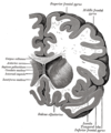
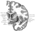
    The caudate nuclei are located near the center of the brain, sitting astride the thalamus. There is a caudate nucleus within each hemisphere of the brain. Individually, they resemble a C-shape structure with a wider "head" (caput in Latin) at the front, tapering to a "body" (corpus) and a "tail" (cauda). Sometimes a part of the caudate nucleus is referred to as the "knee" (genu).[1]


    The head and body of the caudate nucleus form part of the floor of the anterior horn of the lateral ventricle. After the body travels briefly towards the back of the head, the tail curves back toward the anterior, forming the roof of the inferior horn of the lateral ventricle. This means that a coronal (on the same plane as the face) section that cuts through the tail will also cross the body and head of the caudate nucleus.
    The caudate nucleus is related anatomically to a number of other structures. It is separated from the lenticular nucleus (made up of the globus pallidus and the putamen) by the anterior limb of the internal capsule. Together the caudate and putamen form the dorsal striatum.


    Neurochemistry


    The caudate nucleus is highly innervated by dopamine neurons. These neurons originate mainly from the ventral tegmental area (VTA) and the substantia nigra pars compacta (SNc). There are also additional inputs from various association cortices.


    Physiology


    Learning and memory


    Historically, the basal ganglia as a whole have been implicated in higher-order motor control.[2] The caudate nucleus was initially thought to primarily be involved with control of voluntary movement. More recently, it has been demonstrated that the caudate is highly involved in learning and memory,[3] particularly regarding feedback processing.[4] In general, it has been demonstrated that neural activity will be present within the caudate while an individual is receiving feedback. People with "superior autobiographical memory" appear to have slight increases in the sizes of the caudate nucleus as well as of the temporal lobe of the cortex.[5]


     Language comprehension


    The left caudate in particular has been suggested to have a relationship with the thalamus that governs the comprehension and articulation of words as they are switched between languages.[6][7]


    Threshold control


    The brain contains large collections of neurons reciprocally connected by excitatory synapses, thus forming large network of elements with positive feedback. It is difficult to see how such a system can operate without some mechanism to prevent explosive activation. There is some indirect evidence[8] that the caudate may perform this regulatory role by measuring the general activity of cerebral cortex and controlling the threshold potential.


    Role in obsessive compulsive disorder


    It has been theorized that the caudate nucleus may be dysfunctional in persons with obsessive compulsive disorder (OCD), in that it may perhaps be unable to properly regulate the transmission of information regarding worrying events or ideas between the thalamus and the orbitofrontal cortex.
    A neuroimaging study with positron emission tomography found that the right caudate nucleus had the largest change in glucose metabolism after patients had been treated with paroxetine.[9] Recent meta-analyses of voxel-based morphometry studies comparing people with OCD and healthy controls have found people with OCD to have increased grey matter volumes in bilateral lenticular nuclei, extending to the caudate nuclei, while decreased grey matter volumes in bilateral dorsal medial frontal/anterior cingulate gyri.[10][11] These findings contrast with those in people with other anxiety disorders, who evince decreased (rather than increased) grey matter volumes in bilateral lenticular / caudate nuclei, while also decreased grey matter volumes in bilateral dorsal medial frontal/anterior cingulate gyri.[11]


    Additional images


    Thursday, January 6, 2011

    Bushmeat Project; Save the Great Apes


    Say Hi to Brandon
    Make a Chimp Smile
     
    Support Great Ape Protection Act, H.R. 1326, S. 3694
    Please, Sign the Petition


    Bushmeat Project


    Hunting/Bushmeat Bibliography - Persons concerned about great apes and other endangered animals are invited to use and contribute to our working bibliography on hunting and bushmeat commerce in West & Central Africa. Dr. Anthony Rose is a founder and former steering committee member of the Bushmeat Crisis Task Force in Washington, D.C. Read about current BCTF endeavors in North America and Africa."


    Great apes -- gorillas, chimpanzees, and bonobos -- are being hunted to extinction for commercial bushmeat in the equatorial forests of west and central Africa. A ragged far flung army of a few thousand commercial bushmeat hunters supported by the timber industry infrastructure will illegally shoot and butcher more than two billion dollars worth of wildlife this year, including as many as 8,000 endangered great apes. People pay a premium to eat more great apes each year than are now kept in all the zoos and laboratories of the world. If the slaughter continues at its current pace, the remaining wild apes in Africa will be gone within the next fifteen to fifty years. With them will vanish most of the equatorial rain forest, and the cultures of indigenous people who have lived there for millennia.

    It is time that those of us who care about the survival and well-being of the apes, and all life in Africa, confront this crisis. Since our first reports to the international conservation community in 1996 the Bushmeat Project has been urging conservation donors to support programs aimed at helping the African people protect the apes and other endangered animals. The largest wildlife and animal welfare organizations in North America joined us in 1999 in agreement that the Bushmeat Crisis is a top priority concern. It is time to act.

    If we are to stop the slaughter of protected and endangered species we must do so with and through the people who are now involved in the trade, from lorry driver to logging executive, hunter to housewife, gendarme to gentry. Conservation must pursue the biosynergy of humanity and nature in order to find alternative ways to satisfy the human needs that drive the destructive commercial trade in wildlife bushmeat.

    The Bushmeat Project has been established to support partnerships that will help the people of equatorial Africa to protect the region’s vital ecosystems and vibrant societies. The program is a long-term effort to provide economic and social incentive and to enable the expansion of capacity in the conservation arena. For more organizational and program details about the Bushmeat Project please read our Action Agenda, as well as the Project's Mission, Goals and Driving Principles. If you want to join this effort please contact us at bushmeat@biosynergy.org.

    A primary theme of the Bushmeat Project has been the attempt to convert “poachers to protectors.” It began in 1996 when Anthony Rose was introduced to an ex-gorilla hunter in Cameroon’s Eastern Province. See the article "Finding Paradise in a Hunting Camp" to learn how Dr. Rose's involvement in this effort began. The Bushmeat Project has been helping to support the ex-hunter Joseph Melloh in his efforts to become a conservationist. The program led to the establishment of a protected area where Joseph and a small team of conservationists helped two village communities to manage a forest, with the intention of developing gorilla research and tourism as an alternative to hunting and logging. A report on Joseph’s progress in this project and in his other conservation efforts will be posted here soon.

    The biggest challenge now facing conservationists is to reduce the demand for bushmeat. Our Conservation Values Education Program is beginning its fourth year, with expectations for continued success evoking empathy for apes and other primates that will keep people from buying and eating them. Ten thousand copies of Koko's Kitten in French and English were sent to Africa in 2000 and have been used in teaching curricula and community meetings with over three thousand people in schools, training workshops, and rural villages in Cameroon and across the region. Villagers hunters and elders have begun to integrate their own legends about apes and monkeys into the values education curricula, and are taking on the conservation education mission. Results of CVE research in eight villages are being integrated into a major report on this effort.

    The Bushmeat Project and the Biosynergy Institute are working now to build partnerships with other wildlife conservation organizations. The institutionalization and expansion of the Bushmeat Project discoveries and innovations requires an infusion of new organizational structure, as well as enhanced capacity and fund raising processes. Individuals and corporate leaders who have such resources to contribute are invited to contact us at bushmeat@biosynergy.org to discuss your potential for involvement.

    Sunday, January 2, 2011

    Short Description of Tau Protein


    Retrieved From:


    Tau protein


    Microtubule-associated protein tau
    Identifiers
    Symbols MAPT; DDPAC; FLJ31424; FTDP-17; MAPTL; MGC138549; MSTD; MTBT1; MTBT2; PPND; TAU
    External IDs OMIM157140 HomoloGene44834 GeneCards: MAPT Gene
    RNA expression pattern
    PBB GE MAPT 203929 s at tn.png
    PBB GE MAPT 203928 x at tn.png
    PBB GE MAPT 203930 s at tn.png
    More reference expression data
    Orthologs
    Species Human Mouse
    Entrez 4137 17762
    Ensembl ENSG00000186868 ENSMUSG00000018411
    UniProt P10636 P10637
    RefSeq (mRNA) NM_005910 NM_001038609.1
    RefSeq (protein) NP_005901 NP_001033698.1
    Location (UCSC) Chr 17:
    41.33 - 41.46 Mb
    Chr 11:
    103.99 - 104.19 Mb

    PubMed search [1] [2]


    Tau proteins


    They are the proteins that stabilize microtubules. They are abundant in neurons in the central nervous system and are less common elsewhere. When tau proteins are defective, and no longer stabilize microtubules properly, they can result in dementias, such as Alzheimer's disease.
    The tau proteins are the product of alternative splicing from a single gene that in humans is designated MAPT.[1][2] They were discovered in 1975 in Marc Kirschner's laboratory at Princeton University.[3]

     Function


    Tau proteins interact with tubulin to stabilize microtubules and promote tubulin assembly into microtubules. Tau has two ways of controlling microtubule stability: isoforms and phosphorylation. Six tau isoforms exist in brain tissue, and they are distinguished by their number of binding domains. Three isoforms have three binding domains and the other three have four binding domains. The binding domains are located in the carboxy-terminus of the protein and are positively-charged (allowing it to bind to the negatively-charged microtubule). The isoforms with four binding domains are better at stabilizing microtubules than those with three binding domains. The isoforms are a result of alternative splicing in exons 2, 3, and 10 of the tau gene.


    Phosphorylation of tau is regulated by a host of kinases, including PKN, a serine/threonine kinase. When PKN is activated, it phosphorylates tau, resulting in disruption of microtubule organization.[4]Tau protein is a highly soluble microtubule-associated protein (MAP). In humans, these proteins are mostly found in neurons compared to non-neuronal cells. One of tau's main functions is to modulate the stability of axonal microtubules. Tau is not present in dendrites and is active primarily in the distal portions of axons where it provides microtubule stabilization but also flexibility as needed. This contrasts with STOP proteins in the proximal portions of axons which essentially lock down the microtubules and MAP2 that stabilizes microtubules in dendrites. The tau gene locates on chromosome 17q21, containing 16 exons. The major tau protein in the human brain is encoded by 11 exons. Exons 2, 3 and 10 are alternatively spliced, allowing six combinations (2310; 2+310; 2+3+10; 2310+; 2+310+; 2+3+10+). Thus, in the human brain, the tau proteins constitute a family of six isoforms with the range from 352-441 amino acids. They differ in either zero, one or two inserts of 29 amino acids at the N-terminal part (exon 2 and 3), and three or four repeat-regions at the C-terminal part exon 10 missing. So, the longest isoform in the CNS has four repeats (R1, R2, R3 and R4) and two inserts (441 amino acids total), while the shortest isoform has three repeats (R1, R3 and R4) and no insert (352 amino acids total).

     Role in disease


    Hyperphosphorylation of the tau protein (tau inclusions, pTau) can result in the self-assembly of tangles of paired helical filaments and straight filaments, which are involved in the pathogenesis of Alzheimer's disease and other tauopathies.[5]
    All of the six tau isoforms are present in an often hyperphosphorylated state in paired helical filaments from Alzheimer's disease brain. In other neurodegenerative diseases, the deposition of aggregates enriched in certain tau isoforms has been reported. When misfolded, this otherwise very soluble protein can form extremely insoluble aggregates that contribute to a number of neurodegenerative diseases.